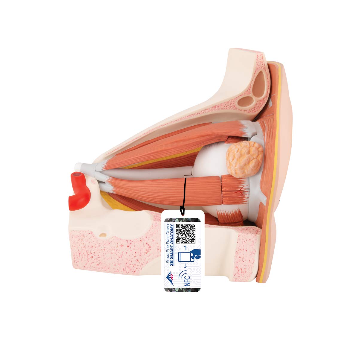
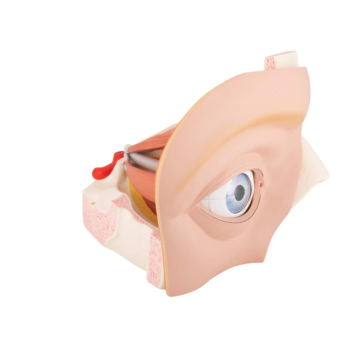
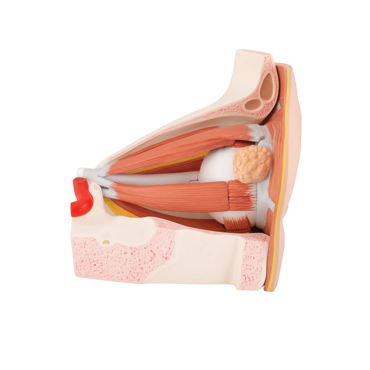
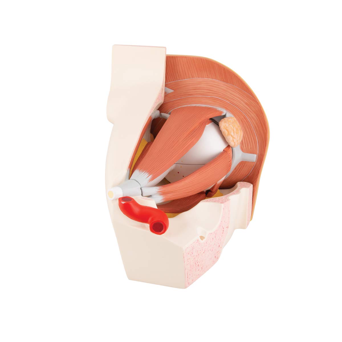
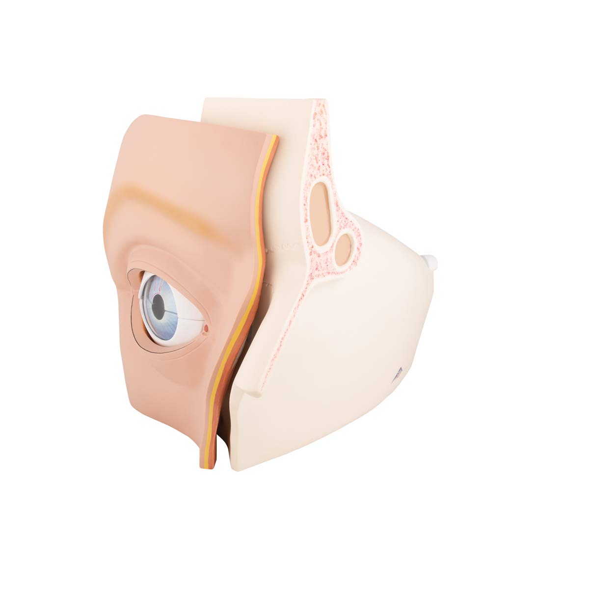
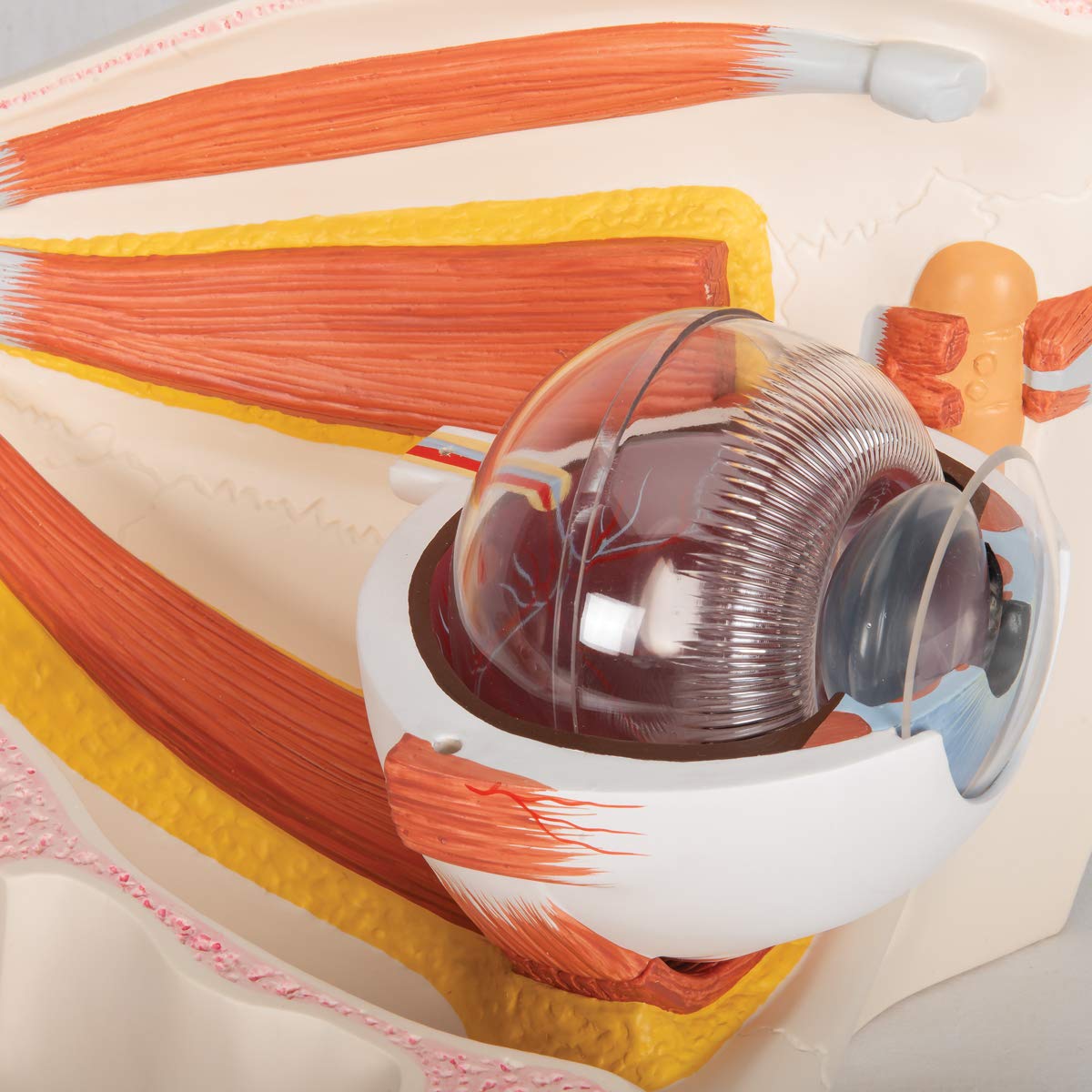
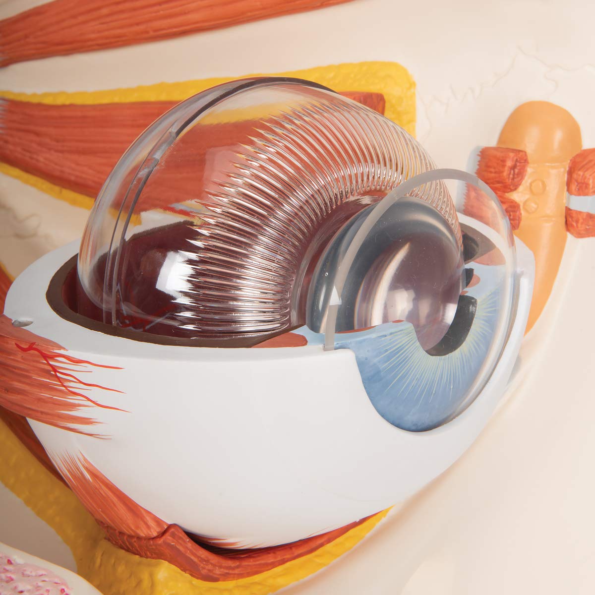
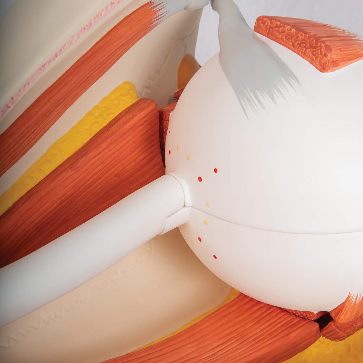
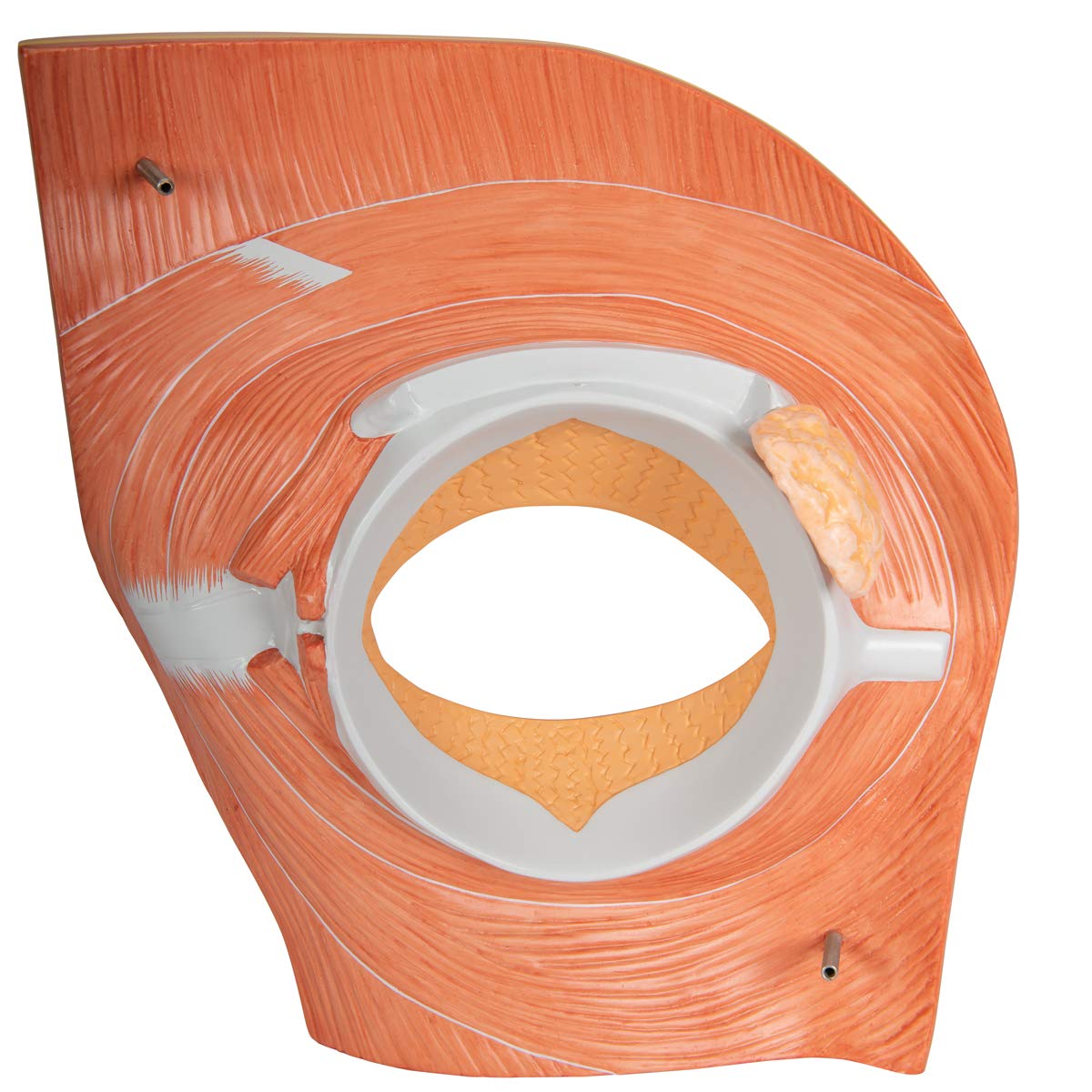
Shows the eyeball with optic nerve in its natural position in the bony orbit (floor and medial wall)
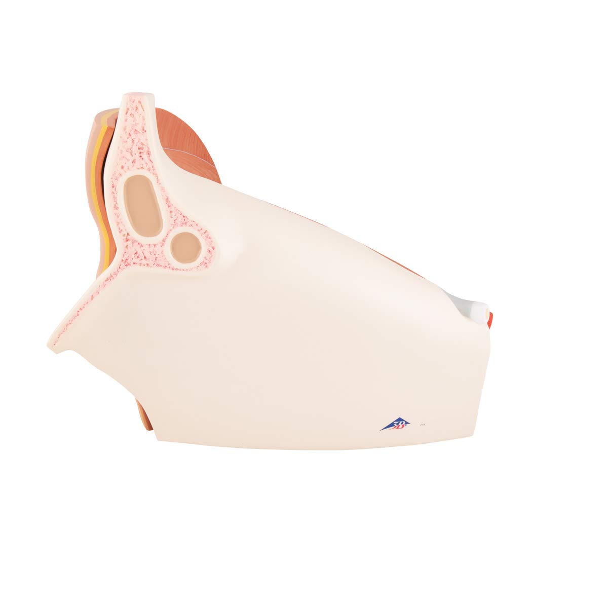
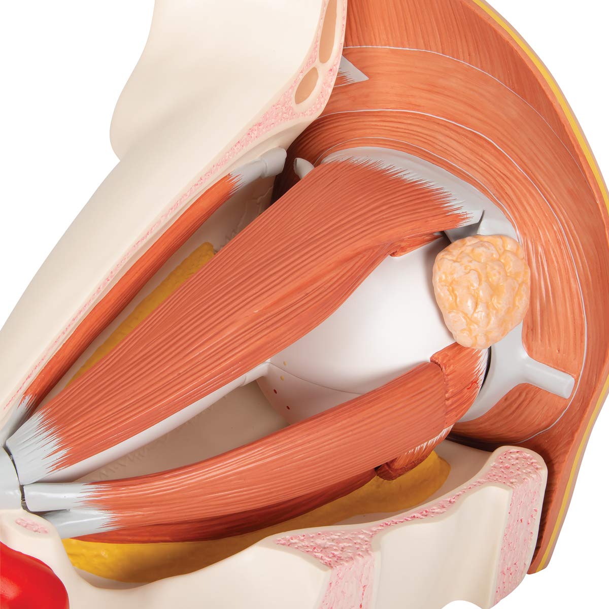
This eye model shows the relation between eye, bones, muscles, and outer structures of the eye
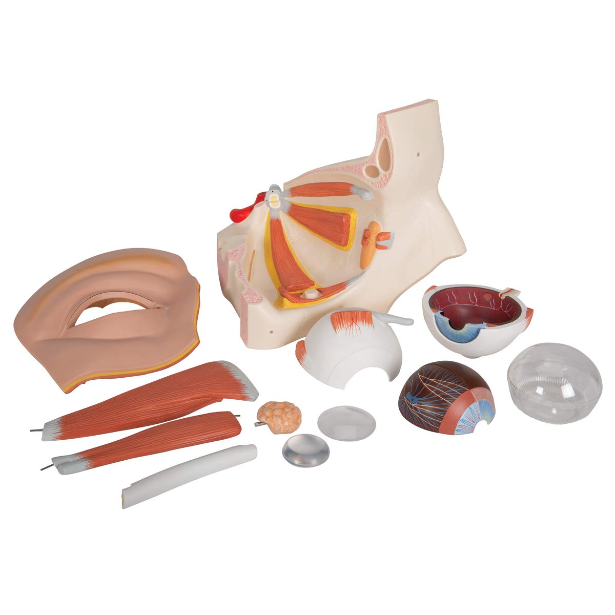
Eye dissects into: Two halves of the sclera, Optic nerve, M. rectus superior, M. rectus lateralis, Cornea half, Eye lens, Lachrymal system, Vitreous humour, Tear gland, Associated structures
Measures: 13" x 11.8" x 15"
Free warranty extension from 3 to 5 years with 3B Smart Anatomy and free access to 3B Smart Anatomy courses in the award-winning Complete Anatomy app. This includes 11 courses with 23 lectures, 117 different views of interactive virtual models, and 39 quizzes.